DONATE NOW
Glaucoma Diagnosis and Optic Nerve Assessment
Your ophthalmologist diagnoses glaucoma by examining the optic nerve and its ability to transmit visual signals to the brain. By assessing optic nerve damage and its progression, your doctor confirms glaucoma or evaluates the stability of the disease. Your ophthalmologist uses various tools, techniques, and instruments to support your care.
Optic Nerve Examination for Glaucoma
Clinicians can readily examine the optic nerve in the clinic using an ophthalmoscope. The optic nerve exits through the back of the eye and contains more than one million individual nerve fibers. These fibers originate in retinal nerve cells, the light-sensitive layer lining the inside of the eye. They travel to various regions of the brain. When your doctor looks into the eye, they see the optic nerve head end-on. The doctor can faintly observe the nerve fibers fanning out across the retina.
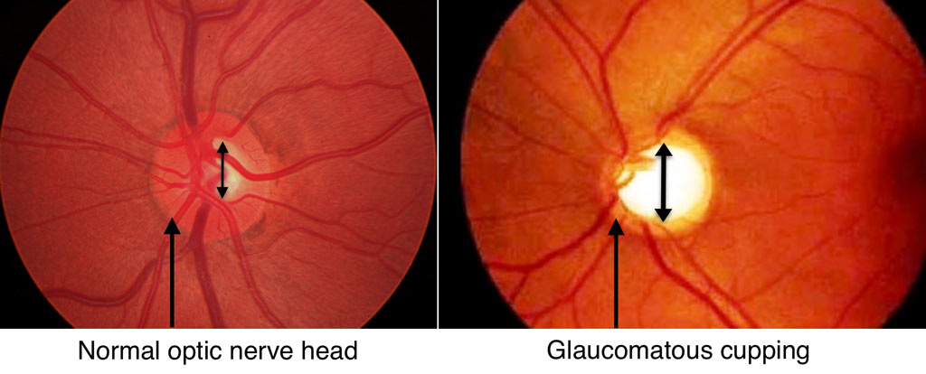
n the normal state, the optic nerve head looks much like a doughnut, with the outer ring consisting of the nerve tissue. Meanwhile, the hole (called the optic cup) is the empty space within the central nerve head that remains after the nerve fibers fan out into the retina. In glaucoma, high eye pressure or other factors damage the nerve fibers, causing them to erode and enlarge the optic nerve cup.
As shown in the optic nerve photograph, a normal optic nerve head has a thick outer ring of nerve tissue with a small optic “cup” centrally. In the glaucomatous optic nerve, the outer ring is thin and the “cup” is larger, corresponding to the loss of nerve fibers. Although many different diseases affect the optic nerve, the damage from glaucoma has a characteristic appearance that permits your ophthalmologist to recognize whether glaucoma is present.
Fundus Photography
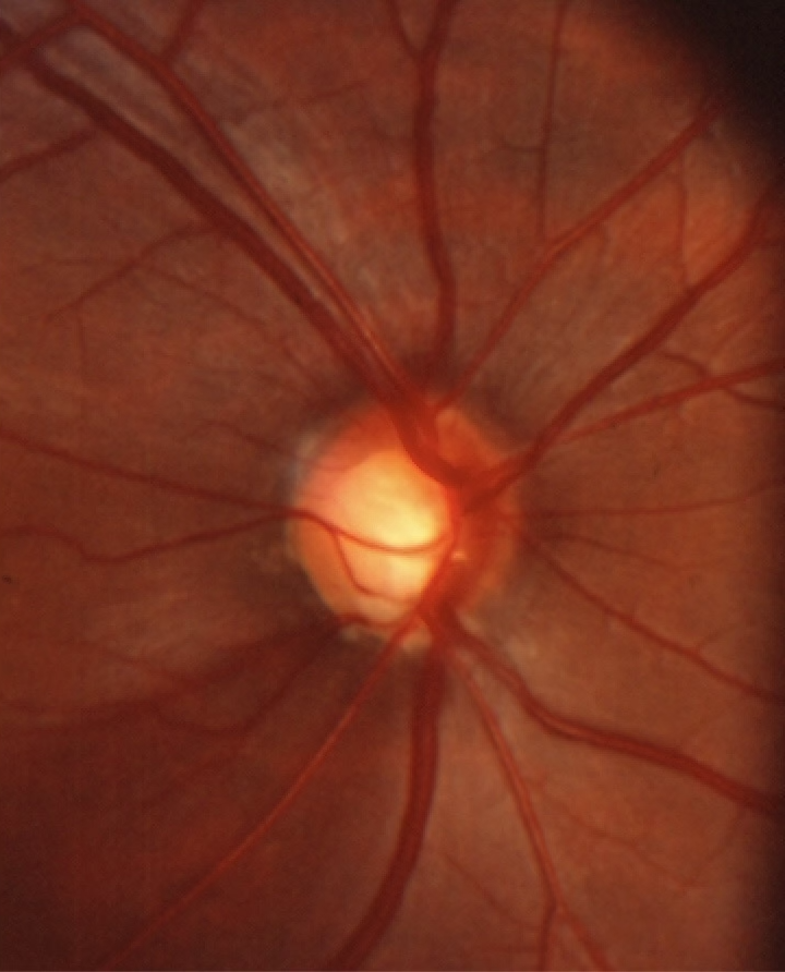
Doctors take fundus photographs to monitor patients suspected of having glaucoma or those already diagnosed. Photographing the optic nerve allows doctors to compare future exams with previous images to track changes.
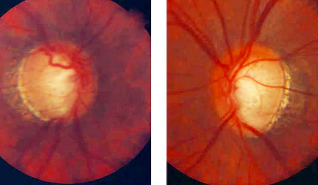
Optic Nerve Changes in Glaucoma
Doctors take fundus photographs to monitor patients suspected of having glaucoma and those diagnosed with the disease. By photographing the optic nerve, doctors can compare future exams with previous images to monitor changes over time. This comparison is invaluable in determining optic nerve progression and deciding on medical and surgical therapy for glaucoma.
Gonioscopy
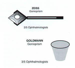
Gonioscopy is the use of a goniolens and a slit lamp or operating microscope to view the anatomical angle formed between the eye’s cornea and iris. Read more about direct and indirect gonioscopy.
Optic Nerve Fiber Analysis
The optic nerve is composed of over one-million individual nerve fibers/axons. Since the axons of nerves cannot be measured accurately in a live eye, indirect measures of axons (axon “counting”) must be used. Optical coherence tomography (OCT), GDx Analyzer (short for Glaucoma Diagnosis Analyzer), and the Heidelberg Retinal Tomography (HRT) are currently used in clinical practice. Currently, OCT is the most popular technology used by glaucoma specialists for optic nerve fiber analysis. Both OCT and GDx are available at Glaucoma Associates of Texas.
Visual Field Testing for Glaucoma
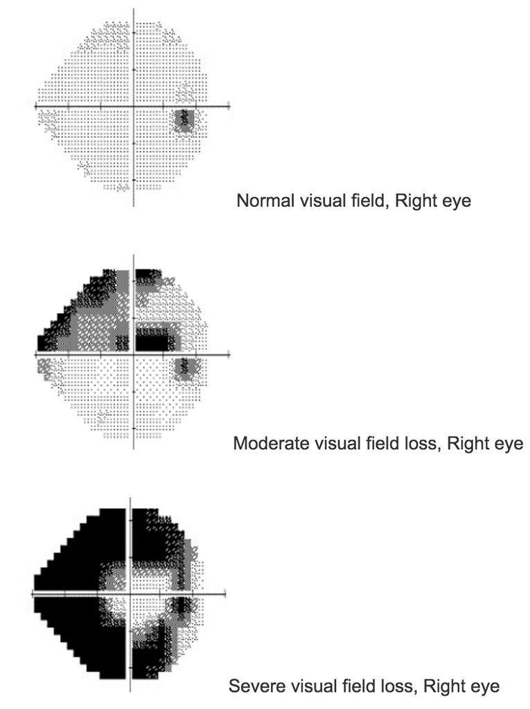
Perhaps the most important tool used to assess optic nerve function, determine whether glaucoma is present, and the status of glaucoma is the visual field test. This test measures how well the optic nerve functions in carrying visual information to the brain. Visual field tests measure the ability of the patient to see light at various points within the retina. Read more about Visual Fields.
Most patients find visual field testing long, tiring, and boring. It is not uncommon to feel that you have performed poorly on a visual field test. Modern visual field machines continue testing each spot measured until the patient misses, in order to determine the dimmest light detectable. Therefore, missing lights can be perfectly normal and should not upset you.