 DONATE NOW
Gonioscopy and Anterior Chamber Angle Evaluation
Gonioscopy refers to the technique used for viewing the drainage system (anterior chamber angle) of the eye for evaluation, classification, and management of normal and abnormal angle structures. The classification of glaucoma relies heavily upon knowledge of the anterior segment anatomy, particularly that of the anterior chamber angle. As a result of their extensive training, all the Doctors at Glaucoma Associates of Texas are experts at gonioscopy and anterior chamber angle evaluation.
Gonioscopy Technique and Equipment
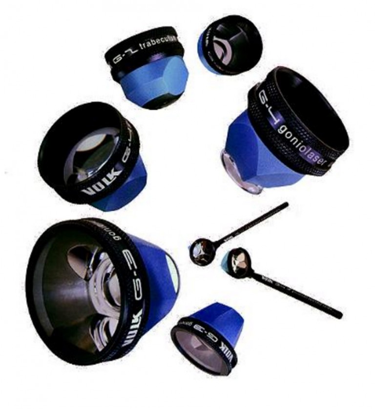
Doctors commonly evaluate the anterior chamber during slit-lamp biomicroscopy, but the chamber angle remains hidden because light rays from it refract back into the eye (total internal reflection). Therefore, doctors use a gonioprism, as shown below.

Types of Gonioprisms
Gonioscopy requires additional effort, skill, and patient co-operation to view the normally concealed chamber angle. Without gonioscopy, it is impossible to classify the type of glaucoma properly.
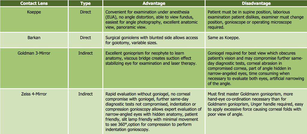
Indirect gonioscopy employs the use of a mirrored goniolens whereas direct gonioscopy does not use a mirror. Most goniolenses used in the clinical setting are indirect goniolenses. The two most common contact lenses for gonioscopy are the Zeiss gonioprism and the Goldman gonioprism.
Gonioscopy Evaluation
During gonioscopy, your doctor will be able to evaluate your anterior chamber angle (drainage system inside the eye). This will allow your doctor to decide whether you have open angle or closed angle glaucoma, or provide clues to other types of glaucoma including Pigment Dispersion Glaucoma or Pseudoexfoliation Glaucoma (see Types of Glaucoma for more information).
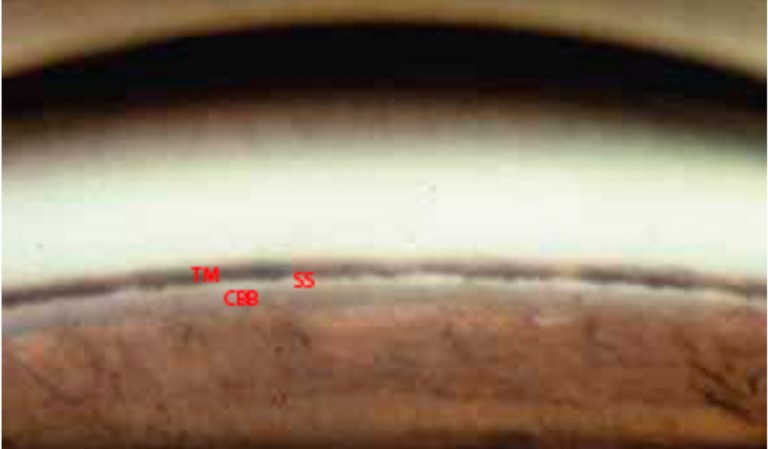
Structures within the anterior chamber angle viewed through a gonioscopy lens. TM: Trabecular Meshwork, SS: Scleral Spur, CBB: Ciliary Body Band)
The Scheie, Shaffer, and Spaeth classifications are the most common grading systems used to classify the appearance of an anterior chamber angle (see below).
Scheie Classification

Shaffer Classification
Spaeth Classification
