DONATE NOW
Glaucoma and Optic Nerve Damage
Glaucoma is a group of eye diseases that cause optic nerve damage due to excessive eye pressure with associated loss of visual function.
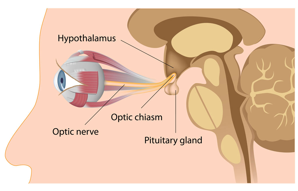
The optic nerve carries visual information from the eye to the brain, much like the cable from a computer screen to the computer.
Glaucoma Definition
The simplest definition of glaucoma is a condition of the eye where the intraocular pressure (pressure inside the eye) is excessive, causing damage to the optic nerve. In the population, average intraocular pressure is 16 mmHg with a range between 8 and 21 mmHg.
When the optic nerve is damaged by excessive pressure, transmission of visual information from the eye to the brain is reduced, causing the visual image and visual field to be impaired.
Fortunately, ophthalmologists can usually detect glaucomatous optic nerve damage before there is noticeable vision loss by examining the back of the eye. Often, damage to the nerve can be detected before there is a change in vision by specialized tests.
Causes of Glaucoma – Optic Nerve and Pressure
In most cases, glaucoma damages the optic nerve due to high eye pressure. Treatment aims to lower eye pressure using medicines, lasers, or surgery. While lost vision cannot be restored, treatment can prevent or slow further loss. In rare cases, the optic nerve continues to deteriorate despite optimal eye pressure. Consequently, researchers worldwide are investigating the causes and developing new treatments to protect the optic nerve.
Many different eye disorders can cause high eye pressure. After measuring the eye pressure, your ophthalmologist attempts to determine the cause of the elevation. It is believed that some form of “clogging” or blockage of the drainage of fluid within the eye (aqueous humor) causes increased eye pressure. Since the eye continually produces aqueous humor, obstruction of its drainage causes the eye pressure to increase. Nearly any eye disorder associated with aging, inflammation, bleeding, injury, tumors, or even birth defects can raise the eye pressure. However, in most cases of glaucoma, examination reveals a normal appearing drainage system and no specific eye abnormalities are found. These patients are described as having “primary open-angle glaucoma.” In other cases, various abnormalities cause a partial or complete blockage of the drainage system. In closed-angle glaucoma, the drainage system is blocked instead of just being clogged.
At least fifty different mechanisms have been described that can raise the eye pressure, but all produce similar damage of the optic nerve.
Ongoing assessment of glaucoma depends upon regular examination of the optic nerve, checking intraocular pressure, and specialized testing.
The well-informed glaucoma patient asks not, “What is my eye pressure, doctor?”, but rather, “How is my optic nerve?”
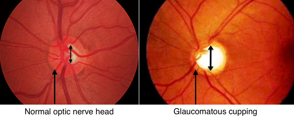
Optic Nerve Cupping in Glaucoma
Damage to the nerve fibers is visible in the appearance of the optic disc. The mechanism(s) by which elevated intraocular pressure eventually results in nerve fiber degeneration is a matter of intense debate.
Microscopic examination of the optic nerve head reveals damage along the length of the nerve fibers and to the mesh-like structure at the optic nerve head that surrounds and supports them.
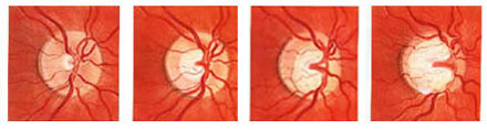
There is a typical sequence of change that occurs to the optic disc in uncontrolled glaucoma.
Optic Nerve Head Cupping (1–4, left to right; C/D = Cup-to-Disc Ratio)
- Normal optic nerve head with small central physiologic cup, C/D ratio ~ 0.2
- Concentric enlargement of the central cup, C/D ratio ~ 0.5
- Irregular enlargement of the cup, especially inferiorly due to loss of inferior neural rim tissue, C/D ratio ~ 0.7
- Marked glaucoma cupping with high degree of central atrophy and loss of inferior rim, C/D ratio ~ 0.9