 DONATE NOW
Detecting Glaucoma Early
A visual field test checks side (peripheral) vision to see if glaucoma has caused damage. Many people have glaucoma without knowing it. In most cases, vision changes happen gradually and without pain.
Glaucoma first affects peripheral vision, which often goes unnoticed. If untreated, it can progress to central vision, making vision loss more obvious. Usually, people notice vision loss only in the later stages.
The test maps vision like a chart. Areas seen well appear light, while areas seen poorly appear dark. This helps doctors detect glaucoma before patients notice changes.
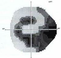
An example of a visual field examination, in an eye with glaucoma. This test measures peripheral (side) vision or visual field. With this test, the visual field looks like a map. Light areas show where vision is strongest. Dark areas show where vision is weak.
“Initially, glaucoma affects peripheral (side) vision… if untreated, glaucoma may eventually affect central vision.”
What Is A Visual Field?
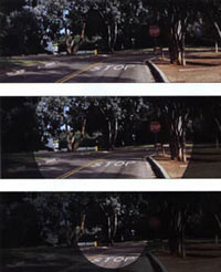
The term visual field refers to the area of a scene you can see with your eyes fixed on one location. For example, while watching a stop light, you may notice a car pulling up beside you in the next lane. Your peripheral vision enables you to see the movement of the car. When both eyes work properly, the visual field covers a wide area (Figure 2). Glaucoma narrows this field of view.
How Doctors Measure the Visual Field
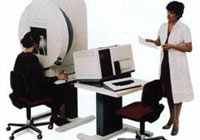
Most ophthalmologists use a device called a perimeter to measure the visual field. During the test, you sit with your head in a chin rest to keep it steady. The examiner asks you to look straight at a small, steady light and keep your eyes still.
While you focus on the light, the examiner flashes small white lights around it – above, below, or to the side – one at a time. Each light corresponds to a spot on your retina. The retina, which lines the inside of the eye, captures light and sends signals through the optic nerve. Glaucoma damages some of these optic nerve fibers.
When both eyes work properly, the visual field covers a wide area (top). As glaucoma worsens, it narrows the field of view (middle) until you see as if you are looking through a tunnel (bottom).
When you see a light flash, you press a button to signal the computer. A computer keeps track of your responses and adjusts the flashes by making them brighter or dimmer at each location tested in the visual field.
The visual field test determines how bright a flashing light must be for you to see it at each location. Every test includes some flashes too dim for even normal eyes to detect. Doctors compare your results to those from healthy eyes. In glaucoma, your eyes detect flashes less easily in certain areas, so the lights must be brighter before you can see them.
“In every test, there will always be some flashes which are too dim for even normal eyes to see.”
Advice For Taking A Visual Field Test
There are several things you can do to be sure you are giving the most accurate responses to the test:
- Get plenty of rest before the test. If you feel tired or sick, ask your ophthalmologist whether to postpone it.
- Sit as comfortably as possible at the perimeter. The tests often take 15 minutes to complete in each eye; so comfort is important. Let the person giving the test know if you feel hunched over or if the chin rest is too high.
- You should always look at the target light. The location of the test lights during the test corresponds to locations on your retina. If you do not look around, your ophthalmologist can more accurately determine from the visual field where damage has occurred.
- You should let the technician know if you need to take a break in order to scratch your nose, cough, or take a rest.
- Do not be concerned if you do not see all the lights. With every test, there will be lights that even someone with normal vision will not be able to see.
What Does The Visual Field Test Tell Your Ophthalmologist?
When your ophthalmologist examines the visual field map, they look for areas less sensitive to light than a healthy eye (Figure 4). Glaucoma often affects certain parts of the visual field more than others (Figure 5). These can help your ophthalmologist to diagnose glaucoma and start early treatment.
If you have glaucoma and have lost some visual field, your doctor will monitor you closely with repeated tests to see if the loss has stopped or is continuing. If your visual field worsens, you may need additional treatment. Your ophthalmologist decides how often to schedule tests based on many factors.
This ongoing and repeated testing is very important for the proper management of your glaucoma.
In a healthy eye, the central portion of the visual field corresponds to the area of best vision. On the left, the numbers show how bright the test lights needed to be for you to detect them. Higher numbers indicate better vision. On the right, the visual field map shows the lightest areas (best vision) are in the center, with some reduction in sensitivity in the periphery, even in the normal field. The darkest area corresponds to the normal blind spot.
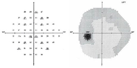
The glaucoma eye shown here required more light than normal in all areas of the field. The numbers on the top are lower, indicating poorer vision. The visual field map (bottom) is much darker than normal, especially in the peripheral areas.