 DONATE NOW
Catalyst For a Cure Vision Restoration Initiative
The Catalyst for a Cure Vision Restoration Initiative, founded by the Glaucoma Research Foundation, brought together four scientists from prestigious academic centers chosen for their expertise in retinal ganglion cell restoration, replacement or repair, neuroprotection, and clinical ophthalmology.
When you donate to the Cure Glaucoma Foundation, the organization uses some of your funds to support this important research program. They pursue their goal of restoring vision lost to glaucoma by developing ways to repair, rebuild, and regenerate the optic nerve. Once successful, the impact of the work by Catalyst for a Cure will be profound as currently there are no effective therapies to restore vision.
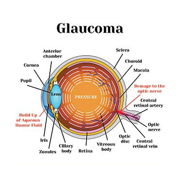
There are over a million retinal ganglion cells (RGCs) in the human retina, and they allow you to see as they send the image to your brain. Unfortunately, glaucoma kills RGCs, and the body cannot replace them, which leads to vision loss. In addition to creating specialized biological tools to identify the different types of RGCs, the team is developing a method for transplanting RGCs and examining the role of genetic signals that influence RGC development and survival.
(CFC) Vision Restoration Initiative
The Catalyst for the Cure (CFC) Vision Restoration Initiative is pursuing two major goals:
- Developing a strategy for optic nerve cell transplantation
- Developing neuroprotective therapies for glaucoma.
The team hopes to use these therapies to help injured – but not dead – optic nerve cells. They aim to improve transplanted cell survival and halt glaucoma-related vision loss.
In the past year, there has been significant progress on both fronts. A major barrier to optic nerve cell transplantation is the inner limiting membrane (ILM). The ILM blocks injected cells from moving from the vitreous into the retina, where they must connect with other retinal cells. The CFC team developed a surgical technique to remove part of the ILM and is testing whether this improves optic nerve cell transplantation. The team also developed an imaging technique to check whether transplanted cells form useful connections with the retina.
Discovering Neuroprotective Targets for Glaucoma
The team conducted a high-throughput screening looking for chemicals that increase survival and increase (not decrease) connectivity with other nerve cells. Researchers then used artificial intelligence to mine the results and identify a potential drug target called “GCK-IV kinases.” The team showed that strategies that target GCK-IV kinases protect optic nerve cells in the retina as well as those derived from stem cells, improving prospects for transplantation. The researchers published these results in the December 2020 issue of Proceedings of the National Academy of Sciences.
In order to identify novel neuroprotective targets for glaucoma, the team measured the change in expression of every gene in the genome. The CFC collaboration is using molecular scissors called CRISPRs in order to delete candidate genes and test their role in a model of glaucoma developed by one of the investigators.
Identifying Protective Factors and Biomarkers for Optic Nerve Cell Survival
Finally, researchers know that optic nerve cells are not all the same and that subtypes exist. Using the same glaucoma model, CFC investigators found that some subtypes resist glaucoma while others show greater sensitivity. This seemed to correlate with the expression of a protein called osteopontin (i.e. more resistant cells had higher levels). The team added osteopontin to the retina. This made previously susceptible optic nerve cells resistant, confirming its protective role. Conversely, if they removed osteopontin, resistant cells became more sensitive. Finally, they showed that osteopontin might be a potential biomarker of glaucoma.
Taken together, the team has several exciting candidate targets to improve optic nerve cell survival. The CFC team plans to translate these techniques to the clinic. They are also improving optic nerve cell transplantation.
Xin Duan, PhD
Assistant Professor, Department of Ophthalmology and Physiology
Weill Institute for Neurosciences
University of California, San Francisco
Dr. Duan’s laboratory investigates retinal ganglion cells subtype-intrinsic factors and tests their roles in optic nerve regeneration and vision recovery.
Yang Hu, MD, PhD
Assistant Professor, Department of Ophthalmology
Stanford University School of Medicine
Stanford, CA
The Hu laboratory focuses on the mechanisms responsible for neuronal degeneration and axon regeneration while maintaining a consistent focus on clinically relevant scenarios and therapies that will translate into effective vision restoration treatments.
Anna La Torre, PhD
Associate Professor, Department of Cell Biology and Human Anatomy
School of Medicine, University of California, Davis
Davis, CA
Dr. La Torre’s laboratory focuses on generating retinal ganglion cells from stem cells to enhance axonal growth and cell survival and ultimately, to use these cells as donor cells for cell replacement therapies and disease modeling.
Derek Welsbie, MD, PhD
Assistant Professor of Ophthalmology, San Diego Shiley Eye Institute
University of California, San Diego
The Welsbie lab identifies genes that cause retinal ganglion cell death, degeneration, and regeneration, and develops new neuroprotective drug therapies for these cells.