
 DONATE NOW
Valve-Like Structures in the Eye’s Drainage System
Valve-like structures discovered in the drainage system of the living eye.
Researchers at Glaucoma Associates of Texas in collaboration with scientists from around the world, determined how to image tiny valve-like structures in the drainage system of the living human eye. These structures were noted in laboratory work from cadaver eyes for decades, but were unable to be visualized in the clinic, until our team determined how to image the tiny valve-like structures, and our patients were a vital part of that team.
Much of the ongoing research, now on three continents, is based on the discovery of these iVaLS, recently named by our team, which stands for intracanalicular Valve-Like Structures. The identification of these iVaLS and their associated links is stimulating research around the world, with many more publications to come. Understanding these valve-like structures in the drainage system of the eye may lead to a better understanding of why the eye pressure becomes abnormal in glaucoma.
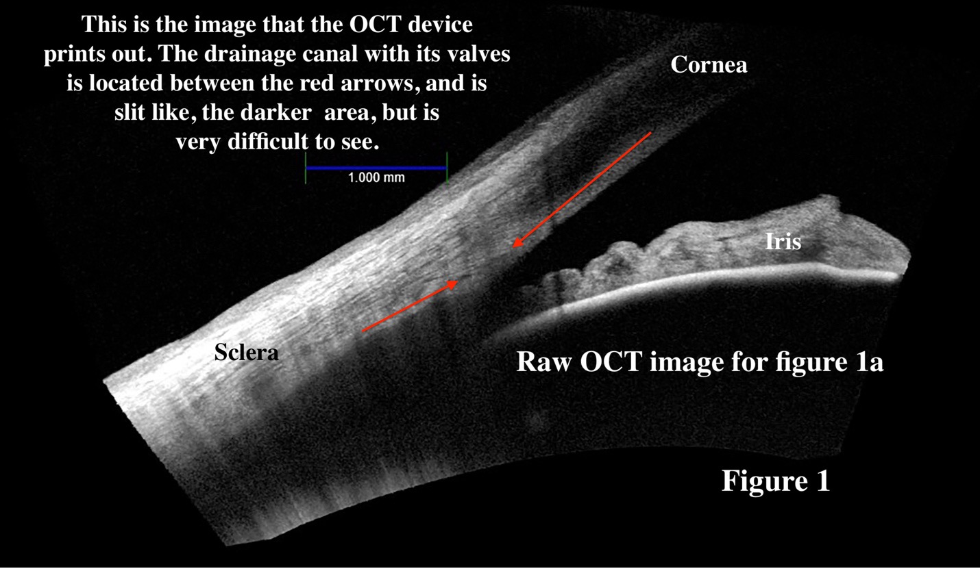
The original publication, which has images from our patients, is posted for your review (article link). The heart of the article is based on reviewing the anatomic structures from the laboratory and comparing them to the images from our patients eyes. For example, take a look at this image from our clinic, it is difficult to find the valve-like structure in the drainage canal (Fig 1).
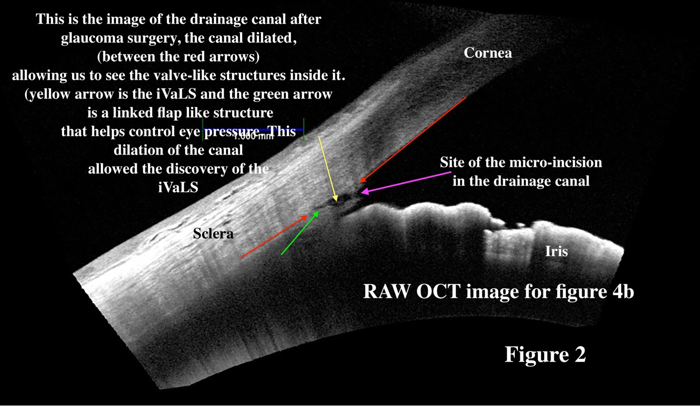
After glaucoma surgery, when the drainage canal dilated to allow more flow into the drain which lowers eye pressure, it was easier to see structures in the drainage canal (Fig 2). Again, the drainage canal is located between the two red arrows and is much easier to see. We compared these clinic images to the laboratory images, and found similarities, the basis for the publication.
For more information on the iVaLS, see the full article, but the above is a simple summary.