 DONATE NOW
What is Cataract Surgery?
Your doctor might recommend cataract surgery or glaucoma surgery to treat your glaucoma. The management approach in the patient who has both glaucoma and cataract must be in many ways totally different from that of a person with cataract uncomplicated by glaucoma. Surgical techniques for both diseases have improved greatly and surgeons with up-to-date expertise in both subspecialties can provide superior outcomes for their patients. The doctors at Glaucoma Associates of Texas are expert cataract surgeons, with particular expertise in complex cataract surgery in glaucoma patients.
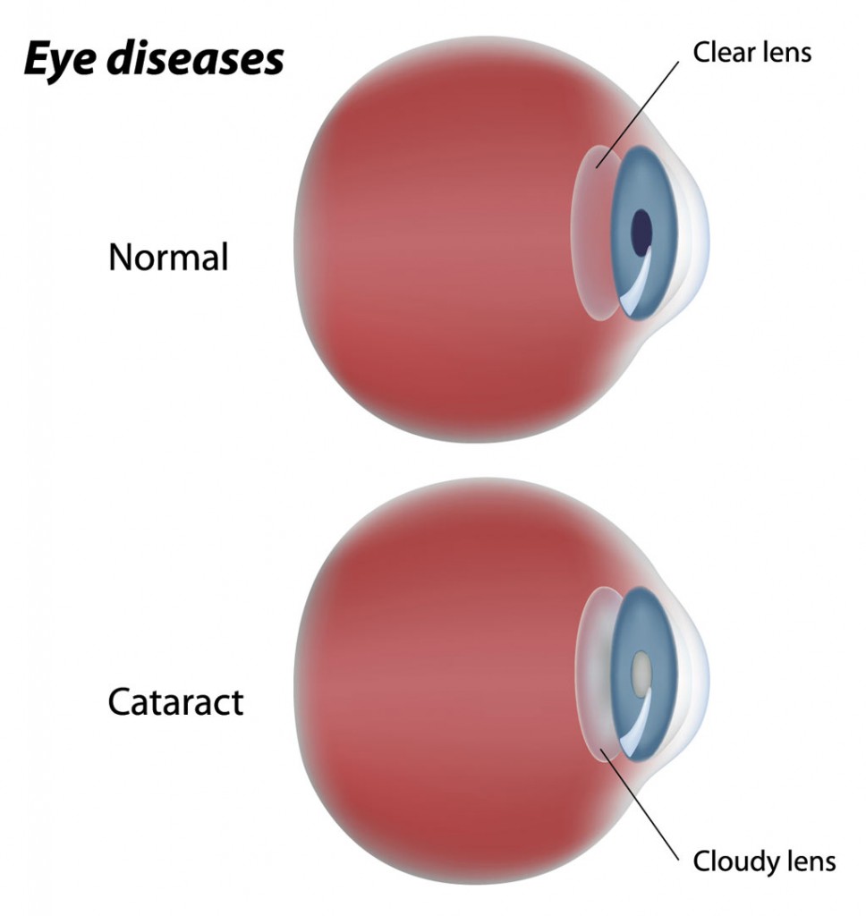
What is a Cataract?
A cataract is hardening and clouding of the natural crystalline lens within your eyeball. It is a normal process of aging. However, this process can occur sooner in glaucoma patients secondary to chronic use of glaucoma drops or from previous glaucoma surgery. In an eye with a narrow angle, a cataract further narrows the angle recess. In such eyes removal of the cataract alone will widen the anterior chamber angle and help prevent an acute angle closure attack.
Your doctor might recommend cataract surgery for treatment of your glaucoma and improvement of your vision. Often this may be combined with a glaucoma procedure as well, such as a GATT, trabeculectomy, glaucoma drainage implant, or others.
Cataract Surgery Overview
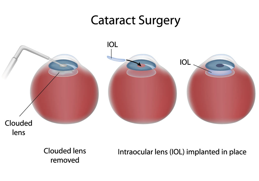
Removal of the cataract is a surgical procedure. Modern day cataract surgery is small incision and often sutureless. Your surgeon makes a tiny incision in the clear part of your eye, called the cornea. They create a circular opening in the capsule that holds the cataract. Next, they insert a probe through this incision. The surgeon breaks the cataract into tiny pieces using an ultrasound probe and removes the debris with an automated irrigation-aspiration system. After clearing all the lens material, they place an intraocular lens implant (IOL) into the capsule. The artificial lens sits in its natural position and remains there for life. If the capsular bag is unstable (which is more common is some glaucoma patients), your doctor may chose to implant the IOL in a different position felt to be safer. After completing the procedure, your doctor may place a stitch in your corneal wound, especially if combined with glaucoma surgery. They remove this stitch easily in the clinic without causing any discomfort.
What Are My Lens Implant Choices?
Before cataract surgery, your doctor measures your eye to determine the correct IOL power. They measure the eyeball’s axial length and corneal curvature. Based on these measurements, your doctor presents various IOL options. In general, there are 3 types of IOL choices:
- Monofocal lens implant: This is the “standard” lens that is covered by insurance without additional cost to the patient. It has the ability to correct your vision at distance (most commonly preferred by patients) or near, but not both. Often, reading glasses are needed for up-close vision. This lens is not able to correct astigmatism.
- Toric lens implant: This lens is able to decrease or eliminate your astigmatism (astigmatism causes distortion of vision if not corrected). However, similar to a “standard” lens above, it cannot correct both distance and near vision, and most patients will require reading glasses for up-close vision.
- Multifocal lens implant: This lens has the ability to improve BOTH your distance and near vision, decreasing and possibly eliminating the need for glasses.
Depending on the measurements completed prior to surgery, one or more of these IOLs may be options. Your doctor will discuss these options with you prior to surgery.
What Steps Does Cataract Surgery Involve?
When you and your doctor make a decision to proceed with Cataract surgery alone or in combination with a glaucoma procedure, you will meet with our preoperative scheduling nurse who will give you detailed instructions on how to prepare yourself for your upcoming surgery. See Preoperative Instructions for more information.
The surgeon performs cataract surgery as an outpatient procedure in an ambulatory surgery center. The procedure usually takes about 30 minutes. Doctors perform it using eye drops or local anesthesia with intravenous sedation. Rarely, they use general anesthesia, putting the patient to sleep. Local anesthesia offers several advantages: it carries less risk, especially for elderly or medically complex patients, reduces post-surgery pain, avoids a sore throat from the airway tube, and allows patients to regain alertness quickly without nausea.
After surgery, your doctor covers the eye with a patch and protects it with a plastic shield overnight. When you have surgery under topical anesthesia (anesthetic drops or gel), your doctor may only place a plastic shield over your eye and instruct you to start eye drops right away. The morning after surgery, your ophthalmologist removes the eye patch and examines your eye. Then, they start eye drops to prevent infection and reduce inflammation. It is important to take these as directed by your doctor since they can make a great deal of difference in the success of the procedure. See Postoperative Instructions for more details.
It takes about 4 to 6 weeks after cataract surgery for the vision to stabilize. You will be ready to change your glasses prescription (if needed) at that time.